Doctoral Candidate
Cupertino, California
Co-Advisor: Professor Alex Dunn
Education B.S. Chemical Engineering, University of California, Berkeley, 2012.
Lab Duties Lab Biosafety Co-coordinator
Research Focus: Rheotaxis: probe how spatial gradients in shear stress influence endothelial cell function
Endothelial cells (ECs) line the inner surface of blood and lymphatic vessels and are highly sensitive to fluid flow as part of their physiological function. EC organization, migration and the vessel development are profoundly influenced by hydrodynamic stresses, with important implications in cardiovascular disease and tumor metastasis. How endothelial cells (ECs) sense fluid flow is a central and unanswered question in cardiovascular biology.
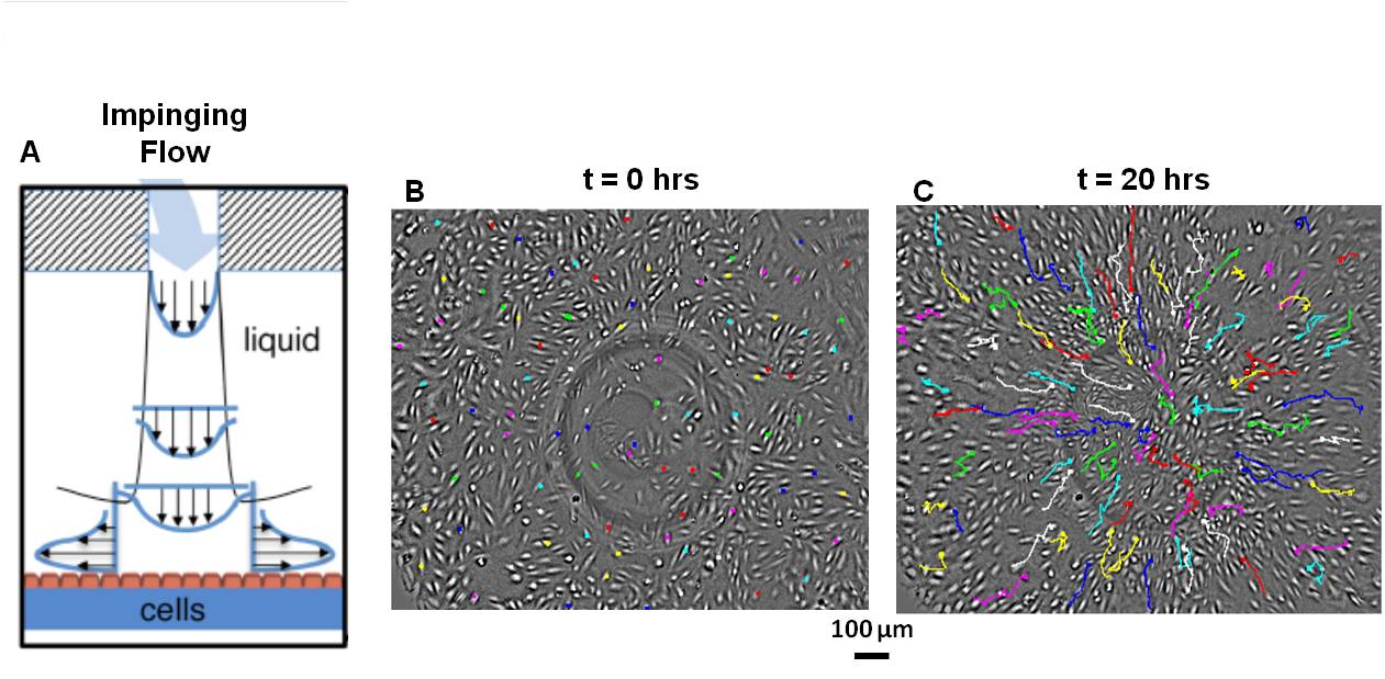
We have developed a high-throughput live-cell fluid flow chamber which models the physiological shear stress gradients experienced by ECs in flow stagnation regions at vessel branches and bifurcations. Live-cell, time-lapse imaging allows us to probe important cellular response to flow, most notably EC migration, which has a key role in vessel remodeling, growth and development. We find that most EC subtypes, including ECs from the venous, arterial, and microvascular systems, migrate in the flow direction. In contrast, human lymphatic microvascular ECs (hLMVECs) migrate against flow and up spatial gradients in wall shear stress, which we have termed rheotaxis1.
Current work aims to both (1) determine the physical underlying mechanism coupling hydrodynamic stresses to chemical signaling in ECs and (2) connect this in a broader scope to vessel development in angiogenesis and lymphangiogenesis, with relevant applications to embryonic development and cardiovascular disease.
Publications:
1. Ostrowski, Maggie A., et al. “Microvascular Endothelial Cells Migrate Upstream and Align Against the Shear Stress Field Created by Impinging Flow.”Biophysical journal 106.2 (2014): 366-374.
Figure 11. (A) Schematic of fluid flow chamber. ECs (orange) are seeded on a gelatin substrate (blue) and are exposed to impinging flow, physiologically relevant in flow stagnation regions. (B) Live-cell imaging allows us to visualize EC migration in response to flow and (C) shows that hLMVECs migrate inward toward jet center against flow direction.